Organisation of cells animal tissues
| Philosophy of Science |
While creating a resource page, please click here for a resource creation checklist
Concept Map
Error: Mind Map file Animal tissues.mm not found
Textbook
Additional information
Useful websites
Reference Books
NCERT Textbook Chapter Tissues
Teaching Outlines
Concept #1 Animal tissues-Epithelial tissue
Learning objectives
- Epithelial tissue perform the function of protection.
- Epithelium covers most organs and cavities within the body.
Notes for teachers
Blood and muscles are both examples of tissues found in our body. On the basis of the functions they perform we can think of four different types of animal tissues, such as epithelial tissue, connective tissue, muscular tissue and nervous tissue. Blood is a type of connective tissue, and muscle forms muscular tissue.
EPITHELIAL TISSUE
The skin, the lining of the mouth, the lining of blood vessels, lung alveoli and kidney tubules are all made ofepithelial tissue. Epithelial tissue cells are
tightly packed without intercellular spaces and form a continuous sheet.
Epithelial tissue is classified into 3types-Squamous epithelium, Columnar epithelium and Cuboidal epithelium.
Squamous epithelium
In cells lining blood vessels or lung alveoli, where transportation of substances occurs through a selectively permeable surface, there is a simple flat kind of epithelium. This is called the simple squamous epithelium. Simple squamous epithelial cells are extremely thin and flat and form a delicate lining.The oesophagus and the lining of the mouth are also covered with squamous epithelium. The skin, which protects the body, is also made of squamous epithelium. Skin epithelial cells are arranged in many layers to prevent wear and tear. Since they are arranged in a pattern of layers,the epithelium is called stratified squamous epithelium.
 Simple squamous epithelium
Where absorption and secretion occur, as in the inner lining of the intestine, tall epithelial cells are present called columnar epithelium. This columnar (meaning ‘pillar-like’) epithelium facilitates movement across the epithelial barrier. In the respiratory tract, the columnar epithelial tissue also has cilia, which are hair-like projections on the outer surfaces of epithelial cells. These cilia can move, and their movement pushes the mucus forward to clear it. This type of epithelium is thus ciliated columnar epithelium.
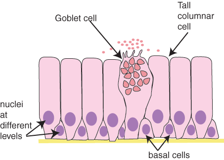 Simple columnar cells
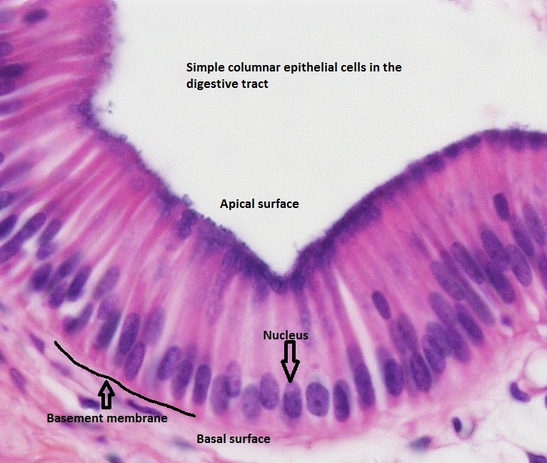 Simple columnar cells in the digestive tract
 ciliated columnar epithelium
Epithelium consisting of cube shaped cells is known as cuboidal epithelium.They form the lining for many ducts such as pancreatic duct, salivary duct and sweat ducts. Epithelial cells often acquire additional specialisation as gland cells, which can secrete substances at the epithelial surface. Sometimes a portion of the epithelial tissue folds inward, and a multicellular gland is formed. This is glandular epithelium.

 Cuboidal epithelium
Activities
- Activity No #1Epithelial_tissue_1
- Activity No #2Epithelial_tissue_2
Concept #2 Muscular tissue
Learning objectives
- Muscular tissue is responsible for the movements of the body.
- The movement of the internal organs like heart,stomach are also caused by muscles.
Notes for teachers
Muscular tissue consists of elongated cells, also called muscle fibres.Muscles contain special proteins called contractile proteins, which contract and relax to cause movement.This property is responsible for movement of limbs and bending of body.The movement of internal organs like heart, stomach and alimentary canal are also caused by muscles.
Muscles are of three types, based on structure,function and location.They are;
- Striped muscles
- Unstriped muscles
- Cardiac muscles
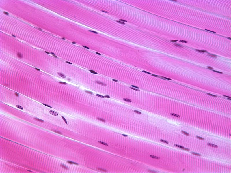
Striped muscles
Muscles present in our limbs can be moved or stopped by our conscious will. Such muscles are called voluntary muscles. These muscles are also called
skeletal muscles as they are mostly attached to bones and help in body movement. Under the microscope, these muscles show alternatelight and dark bands or striations when stained appropriately. As a result, they are also called striated muscles. The cells of this tissue are long, cylindrical, unbranched and
multinucleate.These muscles fatigue easily.
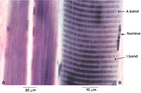 Longitudinal section of Striped muscles
 T.S of Striped muscles
Unstriped muscles
Unstriped muscles are made up of spindle shaped, elongated muscle fibres without striations. They are called smooth muscles.The movement of food in the alimentary canal or the contraction and relaxation of blood vessels are involuntary movements. We cannot control these movements on our own will. Smooth muscles or involuntary muscles control such movements. They are also found in the iris of the eye, in ureters and in the bronchi of the lungs.
 Unstriped muscles
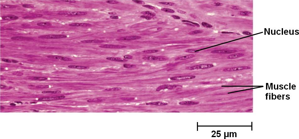 Unstriped muscles
Cardiac muscles
The muscles of the heart show rhythmic contraction and relaxation throughout life. These involuntary muscles are called cardiac muscles. Heart muscle cells are cylindrical,elongated, branched and uninucleate. They do not fatigue easily.
 Cardiac muscles
 Cardiac muscles
 Cardiac muscles
Activities
- Activity No #1Muscular_tissue
- Activity No #2 page_name_concept_name_activity2
Concept #3 Connective tissue
Learning objectives
Notes for teachers
This is the most widespread and abundant type of tissue in the human body. Its function is primarily to support, anchor and connect various parts of the body. Although connective tissue exists in a number of forms, all types have three basic structural elements -- cells, fibres and ground substance. The cells of connective tissue are loosely spaced and embedded in an intercellular matrix . The matrix may be jelly like, fluid, dense or rigid.
Connective tissue is richly supplied with blood.It is also known as binding tissue since it connects or binds other tissues.
Connective tissues are classified into three types on the basis of nature of matrix.
- Loose Connective tissue
- Dense Connective tissue
- Fluid Connective tissue
Loose Connective tissue
The tissue in which the fibres in the matrix are loosely arranged is called loose Connective tissue.Areolar tissue,Adipose tissue and reticular tissue are included under this group.
Areolar tissue
Areolar connective tissue is found between the skin and muscles, around blood vessels and nerves and in the bone marrow. It fills the space inside the organs, supports internal organs and helps in repair of tissues.
The three different fibres of areolar connective tissue are arranged in no particular pattern but run in all directions and form a loose network in the intercellular material.
- Collagen (collagenous) fibres or white fibres are predominant. They usually appear as broad pink bands.
- Yellow fibres or elastic fibres, which appear as thin, dark fibres are also present.
- Reticular fibres.
Adipose tissue
Fat-storing adipose tissue is found below the skin and between internal organs. The cells of this tissue are filled with fat globules. The tissue stores fat which are used as and when the body requires.Storage of fat provides insulation against cold and protects the body like a shock absorber.
Reticular tissue
Activities
- Activity No #1 page_name_concept_name_activity1
- Activity No #2 page_name_concept_name_activity2
Concept #4
Learning objectives
Notes for teachers
Activities
- Activity No #1 page_name_concept_name_activity1
- Activity No #2 page_name_concept_name_activity2